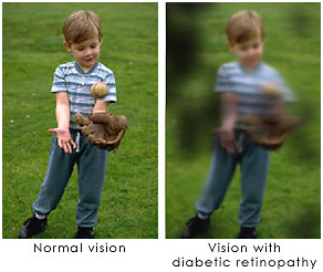
Diabetic retinopathy is a complication of diabetes that is caused by changes in the blood vessels of the retina. When blood vessels in the retina are damaged, they may leak blood and grow fragile, brush-like branches and scar tissue. This can blur or distort the vision images that the retina sends to the brain.
Diabetic retinopathy is a condition occurring in persons with diabetes, which causes progressive damage to the retina, the light sensitive lining at the back of the eye. It is a serious sight-threatening complication of diabetes.
Diabetes is a disease that interferes with the body's ability to use and store sugar, which can cause many health problems. Too much sugar in the blood can cause damage throughout the body, including the eyes. Over time, diabetes affects the circulatory system of the retina.
Apart from the basic eye examination, a thorough fundus examination after dilatation of pupil by indirect ophthalmoscopy is necessary to diagnose diabetic retinopathy. If all the required information cannot be obtained by clinical examination, an investigation called F.F.A (Fundus Fluorescein Angiography) is advised in diabetic patients
In the early stages of diabetic retinopathy, Argon Laser Photocoagulation is used to control the disease and maintain the existing vision. Depending upon the severity of the disease each eye may be given One to Four sittings of Laser treatment.
Diabetic retinopathy is classified into two types : -
Non-proliferative diabetic retinopathy (NPDR) is the early state of the disease in which symptoms will be mild or non-existent. In NPDR, the blood vessels in the retina are weakened causing tiny bulges called microanuerysms to protrude from their walls. The microanuerysms may leak fluid into the retina, which may lead to swelling of the macula.
Proliferative diabetic retinopathy (PDR) is the more advanced form of the disease. At this stage, circulation problems cause the retina to become oxygen deprived.
As a result new fragile blood vessels can begin to grow in the retina and into the vitreous, the gel-like fluid that fills the back of the eye. The new blood vessel may leak blood into the vitreous, clouding vision. Other complications of PDR include detachment of the retina due to scar tissue formation and the development of glaucoma. Glaucoma is an eye disease defined as progressive damage to the optic nerve. In cases of proliferative diabetic retinopathy, the cause of this nerve damage is due to extremely high pressure in the eye. If left untreated, proliferative diabetic retinopathy can cause severe vision loss and even blindness.
Stages of Diabetic Retinopathy
Diabetic retinopathy has four stages : -
Mild Nonproliferative Retinopathy At this earliest stage, microaneurysms occur. They are small areas of balloon-like swelling in the retina's tiny blood vessels.
Moderate Nonproliferative Retinopathy As the disease progresses, some blood vessels that nourish the retina are blocked.
Severe Nonproliferative Retinopathy Many more blood vessels are blocked, depriving several areas of the retina with their blood supply. These areas of the retina send signals to the body to grow new blood vessels for nourishment.
Proliferative Retinopathy At this advanced stage, the signals sent by the retina for nourishment trigger the growth of new blood vessels. This condition is called proliferative retinopathy. These new blood vessels are abnormal and fragile. They grow along the retina and along the surface of the clear, vitreous gel that fills the inside of the eye. By themselves, these blood vessels do not cause symptoms or vision loss. However, they have thin, fragile walls. If they leak blood, severe vision loss and even blindness can result.
Diagnosis
Diabetic retinopathy can be diagnosed through a comprehensive eye examination.
Testing, with special emphasis on evaluation of the retina and macula, may include : -
Patient history to determine vision difficulties experienced by the patient, presence of diabetes, and other general health concerns that may be affecting vision
Visual acuity measurements to determine the extent to which central vision has been affected
Refraction to determine the need for changes in an eyeglass prescription
Evaluation of the ocular structures, including the evaluation of the retina through a dilated pupil
Measurement of the pressure within the eye
Supplemental testing may include : -
Retinal photography or tomography to document current status of the retina
Fluorescein angiography to evaluate abnormal blood vessel growth
Symptoms diabetic retinopathy
Often there are no symptoms in the early stages of the disease, nor is there any pain. Don't wait for symptoms. Be sure to have a comprehensive dilated eye exam at least once a year.
Blurred vision may occur when the macula-the part of the retina that provides sharp central vision-swells from leaking fluid. This condition is called macular edema.
If new blood vessels grow on the surface of the retina, they can bleed into the eye and block vision.
In patients with diabetes, prolonged periods of high blood sugar can lead to the accumulation of fluid in the lens inside the eye that controls eye focusing. This changes the curvature of the lens and results in the development of symptoms of blurred vision. The blurring of distance vision as a result of lens swelling will subside once the blood sugar levels are brought under control. Better control of blood sugar levels in patients with diabetes also slows the onset and progression of diabetic retinopathy.
Often there are no visual symptoms in the early stages of diabetic retinopathy. That is why the American Optometric Association recommends that everyone with diabetes have a comprehensive dilated eye examination once a year. Early detection and treatment can limit the potential for significant vision loss from diabetic retinopathy.
Treatment of diabetic retinopathy varies depending on the extent of the disease. It may require laser surgery to seal leaking blood vessels or to discourage new leaky blood vessels from forming. Injections of medications into the eye may be needed to decrease inflammation or stop the formation of new blood vessels. In more advanced cases, a surgical procedure to remove and replace the gel-like fluid in the back of the eye, called the vitreous, may be needed. A retinal detachment, defined as a separation of the light-receiving lining in the back of the eye, resulting from diabetic retinopathy, may also require surgical repair.
If you are a diabetic, you can help prevent or slow the development of diabetic retinopathy by taking your prescribed medication, sticking to your diet, exercising regularly, controlling high blood pressure and avoiding alcohol and smoking.
Causes of diabetic Retinopathy
Diabetic retinopathy is the result of damage to the tiny blood vessels that nourish the retina. They leak blood and other fluids that cause swelling of retinal tissue and clouding of vision. The condition usually affects both eyes. The longer a person has diabetes, the more likely they will develop diabetic retinopathy. If left untreated, diabetic retinopathy can cause blindness.
Q. What does diabetic Retinopathy causes ?
Blood vessels damaged from diabetic retinopathy can cause vision loss:
Fluid can leak into the macula, the area of the retina which is responsible for clear central vision. Although small, the macula is the part of the retina that allows us to see colors and fine detail. The fluid causes the macula to swell, resulting in blurred vision.
In an attempt to improve blood circulation in the retina, new blood vessels may form on its surface. These fragile, abnormal blood vessels can leak blood into the back of the eye and block vision
Risk factors for diabetic retinopathy include : -
Diabetes : - people with Type 1 or Type 2 diabetes are at risk for the development of diabetic retinopathy. The longer a person has diabetes, the more likely they are to develop diabetic retinopathy, particularly if the diabetes is poorly controlled.
Race: - Hispanic and African Americans are at greater risk for developing diabetic retinopathy.
Medical conditions : - persons with other medical conditions such as high blood pressure and high cholesterol are at greater risk.
Pregnancy : - pregnant women face a higher risk for developing diabetes and diabetic retinopathy. If gestational diabetes develops, the patient is at much higher risk of developing diabetes as they age.
If remains untreated it may lead to severe diseases such as Glaucoma and Ratinal Detachment.
Q. How does diabetes affect the retina ?
Patients with diabetes are more likely to develop eye problems such as cataracts and glaucoma, but the disease's affect on the retina is the main threat to vision. Most patients develop diabetic changes in the retina after approximately 20 years. The effect of diabetes on the eye is called diabetic retinopathy.
Over time, diabetes affects the circulatory system of the retina. The earliest phase of the disease is known as background diabetic retinopathy. In this phase, the arteries in the retina become weakened and leak, forming small, dot-like hemorrhages. These leaking vessels often lead to swelling or edema in the retina and decreased vision.
The next stage is known as proliferative diabetic retinopathy. In this stage, circulation problems cause areas of the retina to become oxygen-deprived or ischemic. New, fragile, vessels develop as the circulatory system attempts to maintain adequate oxygen levels within the retina. This is called neovascularization. Unfortunately, these delicate vessels hemorrhage easily. Blood may leak into the retina and vitreous, causing spots or floaters, along with decreased vision.
In the later phases of the disease, continued abnormal vessel growth and scar tissue may cause serious problems such as retinal detachment and glaucoma.
Q. How is diabetic retinopathy treated ?
Treatment for diabetic retinopathy depends on the stage of the disease and is directed at trying to slow or stop the progression of the disease.
scatter laser treatment may still be possible, depending on the amount of bleeding.
During the first three stages of diabetic retinopathy, no treatment is needed, unless you have macular edema. To prevent progression of diabetic retinopathy, people with diabetes should control their levels of blood sugar, blood pressure, and blood cholesterol.
In the early stages of diabetic retinopathy, Argon Laser Photocoagulation is used to control the disease and maintain the existing vision. Depending upon the severity of the disease each eye may be given One to Four sittings of Laser treatment.
In the early stages of Non-proliferative Diabetic Retinopathy, treatment other than regular monitoring may not be required. Following your doctor's advice for diet and exercise and keeping blood sugar levels well-controlled can help control the progression of the disease.
If the disease advances, leakage of fluid from blood vessels can lead to macular edema. Laser treatment (photocoagulation) is used to stop the leakage of blood and fluid into the retina. A laser beam of light can be used to create small burns in areas of the retina with abnormal blood vessels to try to seal the leaks.
Proliferative retinopathy is treated with laser surgery. This procedure is called scatter laser treatment. Scatter laser treatment helps to shrink the abnormal blood vessels. Your doctor places 1,000 to 2,000 laser burns in the areas of the retina away from the macula, causing the abnormal blood vessels to shrink. Because a high number of laser burns are necessary, two or more sessions usually are required to complete treatment.
Although you may notice some loss of your side vision, scatter laser treatment can save the rest of your sight. Scatter laser treatment may slightly reduce your color vision and night vision.
Scatter laser treatment works better before the fragile, new blood vessels have started to bleed. That is why it is important to have regular, comprehensive dilated eye exams. Even if bleeding has started, If the bleeding is severe, you may need a surgical procedure called a vitrectomy. During a vitrectomy, blood is removed from the center of your eye.
Persons with diabetic retinopathy can suffer significant vision loss. Special low vision devices such as telescopic and microscopic lenses, hand and stand magnifiers, and video magnification systems can be prescribed to make the most of remaining vision.
The list of of Eye Hospitals in India is as follows : -
For more information, medical assessment and medical quote
send your detailed medical history and medical reports
as email attachment to
Email : - info@wecareindia.com
Call: +91 9029304141 (10 am. To 8 pm. IST)
(Only for international patients seeking treatment in India)
For a detailed evaluation send patient’s medical reports / X rays / doctors notes to info@wecareindia.com
Patient Storys
Successful heart surgery at We Care India partner hospital allows Robert Clarke to live a normal life despite a rare genetic disorder We Care india helped Robert find best super specialised surgeon for his rare conditions.