The only certain way to learn whether a breast lump or mammographic abnormality is cancerous is by having a biopsy, a procedure in which tissue is removed by a surgeon or other specialist and examined under a microscope by a pathologist. A pathologist is a doctor who specializes in identifying tissue changes that are characteristic of disease, including cancer.
Tissue samples for biopsy can be obtained by either surgery or needle. The doctor's choice of biopsy technique depends on such things as the nature and location of the lump, as well as the woman's general health.
symptoms
Fever
Pain
Redness or swelling around the incision
Warmth around the incision
Drainage from the incision
Benign breast conditions
If you find changes or something unusual in one of your breasts, it is important to see a doctor or nurse as soon as possible. But keep in mind that most breast changes are not cancer. Just because your doctor wants you to have a biopsy does not mean you have breast cancer: 4 of every 5 biopsy results are not cancer. But the only way to know for sure is to take out and test tissue from the suspicious area of the breast.
Benign (be-nine) or non-cancerous breast conditions are very common and they are never life threatening. The 2 main types are fibrocystic changes and benign breast tumors.
Fibrocystic changes : -
Fibrocystic changes are benign changes in the breast tissue that happen in about half of all women at some time in their lives. This change often happens just before a menstrual period is about to begin. Although this used to be called fibrocystic disease, it is not a disease at all. These changes can cause cysts (fluid-filled sacs) and areas of lumpiness, thickening, or tenderness; nipple discharge; or pain in the breast. If they are painful, cysts can be treated by taking out the fluid with a needle and syringe, but they may fill up again later.
A cyst cannot be diagnosed by physical exam alone, nor can it be diagnosed by a mammogram alone. To be sure that a lump (mass) is really a cyst, the doctor can do either a breast ultrasound or take the fluid out of the cyst with a thin, hollow needle.
A cyst is filled with fluid. If a mass has any solid parts, it is no longer a simple cyst and you may need to have more imaging tests. Some masses can be watched with mammograms, while others may need a biopsy. The size, shape, and edges (margins) of the mass help the doctor figure out whether cancer may be present.
Lumps and areas of thickening caused by fibrocystic changes are almost always harmless. If fibrocystic changes are uncomfortable or painful, doctors may suggest that you avoid caffeine or reduce your salt intake. In severe cases, doctors can prescribe medicines that may help reduce or relieve your symptoms.
Benign breast tumors : -
Benign breast tumors are non-cancerous areas where breast cells have grown abnormally and rapidly, often forming a lump. Unlike cysts, which are filled with fluid, tumors are solid. Benign breast tumors are sometimes uncomfortable, but they are not dangerous and do not spread outside the breast to other organs. Still, some benign breast conditions, such as papillomas and atypical hyperplasia, are important to know about because women with these conditions have a higher risk of developing breast cancer. For more information see our document, Non-Cancerous Breast Conditions.
A biopsy is the only way to find out if a tumor is benign or cancerous. (See the section "Types of biopsy procedures" for more information.) In a biopsy, part of the lump or suspicious area is removed and looked at under a microscope.
If a benign tumor is large, it may change the breast's size and shape. Depending on the size and number of benign tumors, doctors may recommend that it be removed by surgery (excision).
If the benign tumor is growing into the tissue of the milk ducts, it may cause an abnormal discharge from the nipple. In some cases, this can be treated by surgery to remove the tumor.
Other benign breast conditions
Mastitis : - Mastitis is a breast infection that most often affects women who are breast-feeding. The breast may become red, warm, or painful. Mastitis is treated with antibiotics. But if the mastitis does not get better when you take antibiotics, it is important that you let the doctor know right away. Some breast cancers can look like infections.
Fat necrosis : - Fat necrosis sometimes happens when an injury to the breast heals and leaves scar tissue that can feel like a lump. A biopsy can tell if it is cancer or not. Sometimes when the breast is injured, an oil cyst (fluid-filled area) forms instead of scar tissue during healing. Oil cysts can be diagnosed and treated by taking out (aspirating) the fluid.
Duct ectasia : - Duct ectasia is common and most often affects women in their 40s and 50s. Its symptoms are usually a green, black, thick, or sticky discharge from the nipple, and tenderness or redness of the nipple and area around the nipple. Duct ectasia can also cause a hard lump, which is usually biopsied to be sure it is not cancer. Redness that does not improve may need to be biopsied to be sure it is not cancer.
Non-Cancerous Breast Conditions
Non-cancerous breast conditions are breast changes that are not cancer. They are very common and can be found in most women. In fact, most breast changes that are biopsied and looked at under the microscope turn out to be benign (bee-nine). Benign is another word for non-cancerous.
Unlike breast cancers, benign breast conditions are not life-threatening. But sometimes they can cause symptoms that bother you. And certain benign conditions are linked with an increased risk of developing breast cancer. We will cover this in more detail later.
Q. Which Biopsy you would like to go for ?
One Step Biopsy : -
Not too many years ago, all women undergoing surgery for breast symptoms had a one-step procedure: If the surgical biopsy showed cancer, the surgeon performed a mastectomy immediately. The woman went into surgery not knowing if she had cancer or if her breast would be removed.
Some women, nonetheless, prefer a one-step procedure. They have decided beforehand that, if the surgical biopsy and frozen section show cancer, they want to go ahead with surgery, either mastectomy or lumpectomy and axillary dissection (removal of the underarm lymph nodes). If, on the other hand, the lump proves to be benign, the incision will be closed. The procedure will have taken less than an hour, and the woman may go home the same day or the next day.
A one-step procedure avoids the physical and psychological stress, as well as the costs in time and money, of two rounds of surgery and anesthesia--a particularly important consideration for women who are ill or frail. Women who have symptoms of breast cancer can find the wait between biopsy and surgery emotionally draining, and they may be relieved to have a one-step procedure to take care of the problem as quickly as possible.
Two Step Biopsy : -
Today a woman facing biopsy has a broader range of options. In most cases, biopsy and diagnosis will be separated from any further treatment by an interval of several days or weeks. Such a two-step procedure does not harm the patient, and it has several benefits. It allows time for the tissue sample to be examined in detail and, if cancer is found, it gives the woman time to adjust to the diagnosis. She can review her treatment options, seek a second opinion, receive counseling, and arrange her schedule.
No single solution is right for everyone. Each woman should consult with her doctors and her family, weigh the alternatives, and decide what approach is appropriate. Being involved in the decision-making process can give a woman a sense of control over her body and her life.
ways to do a breast biopsy : -
There are several ways to do a breast biopsy. The sample of breast tissue will be looked at under a microscope to check for cancer cells.
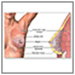
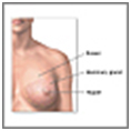
Female Breast
Breast lump removal
fine-needle aspiration biopsy : -
A fine-needle aspiration biopsy puts a thin needle through the skin, into the lump, and removes cells to look at. Needle aspiration may be done to see if the lump is solid or fluid-filled (cyst). If the lump is a cyst, it will go away after the fluid is removed. If there is no fluid, another type of biopsy will be done.
core needle : -
A core needle biopsy uses a large needle fitted with a special tip. The needle goes through the skin to the lump or area to take out a sample of tissue about the size of a pencil lead. A core needle biopsy can also be done using a suction unit that gently removes a larger sample of tissue.
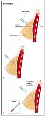
stereotactic biopsy : -
A stereotactic biopsy uses a special type of X-ray during a core needle biopsy to find the area of the breast where the biopsy sample will be taken. This technique can check a lump that cannot be felt on breast examination but is seen on mammogram or ultrasound. A small incision is then made in the skin of the breast, and the core needle is guided by the X-ray to the biopsy site to take a tissue sample. Stereotactic biopsy may not be appropriate for all types of breast lumps.
Open Surgical Breast Biopsy : -
Prior to recent biopsy advancements, physicians and surgeons routinely recommended open surgical breast biopsies. While this remains an accurate procedure, it is the most invasive biopsy procedure and results in external and internal scarring. Most patients recover quickly from a breast biopsy surgery; however, some may experience post-operative pain or minor disfiguring of the breast.
This is considered general surgery, which requires an operating room, general anesthesia in some cases, and stitches. Because of the hospital and surgical resources needed to perform the operation, open surgical biopsies are more costly than other breast biopsy methods. Because most breast biopsies are for small breast abnormalities that cannot be felt (non-palpable lesions), open surgical biopsies are sometimes viewed as excessive.
Open surgical procedures often involve a two-step process. First, a radiologist identifies the area to be biopsied. Through a process known as wire localization, a wire is positioned in the abnormal breast tissue to identify the area to be cut out and removed during the breast biopsy surgery. Next, the patient is taken to the operating room where she is placed under general anesthesia or a local anesthesia with sedation.
A surgeon makes a 1 to 2-inch incision in the breast and removes the localization wire and a large section of tissue, typically about the size of a golf ball. The incision in the breast is then closed with stitches and covered with a protective bandage.
Technique
The patient lies prone on the stereotactic table with the breast suspended through a hole in the table The breast is then placed in compression. Images are then obtained using digital x-rays. These x-rays use much less radiation than traditional mammograms. Images are taken at two 15-degree angles from the center. The images are viewed on a computer monitor, and the physician can identify the lesion in three dimensions.
The computer can then then help guide a biopsy needle to the exact coordinates of the lesion. The breast tissue can be removed in one of two ways. A large bore needle can be used to remove cores of tissue. This is called the Mammotome procedure.
It removes cores of breast tissue via a small incision (2-3mm). Multiple cores are taken (usually 6-10). The major advantage of the Mammotome procedure is that there is virtually no scar. The other type of stereotactic breast biopsy is called the ABBI procedure. This device removes a larger core of tissue (5-20mm).
In this fashion, the entire lesion can be removed. This can sometimes provide a more accurate diagnosis. Both types of stereotactic breast biopsies are performed under local anasthesia. Patients have minimal discomfort during or after the procedure. Patients can usually resume normal activities by the following day. Stereotactic biopsies have been shown to be very accurate. They are as accurate as an open surgical biopsy.
Benefits of the procedure include less patient discomfort, quicker recovery, decreased scarring, and decreased cost. Traditional mammographic directed biopsies require that the lesion be seen on two views, but with stereotactic techniques abnormalities that are seen on one view can be removed. There are certain mammographic lesions that cannot be biopsied stereotactically. These include areas that are vague on the mammogram and might not show up on the digital screen as well as some areas of diffuse calcifications. Technical problems are sometimes seen in patients with small breasts or in lesions that are up against the chest wall.
The decision as to whether a lesion can be removed stereotactically is usually made by the surgeon and the radiologist. As the procedure of stereotactic breast biopsy becomes more popular, more hospitals are obtaining the necessary equipment. Thus the technique is becoming available to the majority of patients with mammographically detectable lesions.
Risks
There is a slight chance of infection at the injection or incision site.
Excessive bleeding is rare, but may require draining or re-bandaging. Bruising is common.
There will be a scar. Depending on the amount of tissue removed and how the breast heals, the appearance of the breast may be affected.
Depending on the results of the biopsy, further surgery or treatment may be needed.
Infection is always a possibility when the skin is broken, although this rarely occurs. Redness, swelling, or severe pain at the biopsy site would indicate a possible infection. Another possible consequence of a breast biopsy is a hematoma. This is a collection of blood at the biopsy site; the body usually absorbs blood naturally.
If the hematoma is very large and uncomfortable, it may need to be drained. A surgical breast biopsy may produce a visible scar on the breast, which may make future mammograms harder to interpret accurately.
A false negative pathology report is another risk. This means that no cancer was found when cancer was actually present. The incidence of this varies with the biopsy technique. In general, fine-needle aspiration biopsies have the highest rate of false negative results, but there may be variation in results between facilities.
FAQs
Q. When can I expect to return to work and/or normal activities ?
Light activity at home is encouraged after surgery. You should be able to return to normal activities shortly after the procedure. If you are taking narcotic medications for pain, you should not drive.
Q. What activities to be done after Biopsy
Avoid strenuous activity, heavy lifting and vigorous exercise until the stitches are removed. Walking is a normal activity that can be restarted right away.
If possible, plan to take the day off or plan a lighter day following the surgery.
Q. How to prepare for the test
Your medical history will be taken, and a manual breast exam performed.
An informed consent form must be signed. If you are going to have general anesthesia, you may be asked to fast for 8 to 12 hours before the test.
If you take medications (including aspirin or herbals), ask your doctor whether to discontinue these before the biopsy.
The list of of General Surgery Hospitals in India is as follows : -
For more information, medical assessment and medical quote
send your detailed medical history and medical reports
as email attachment to
Email : - info@wecareindia.com
Call: +91 9029304141 (10 am. To 8 pm. IST)
(Only for international patients seeking treatment in India)
For a detailed evaluation send patient’s medical reports / X rays / doctors notes to info@wecareindia.com
Patient Storys
Successful heart surgery at We Care India partner hospital allows Robert Clarke to live a normal life despite a rare genetic disorder We Care india helped Robert find best super specialised surgeon for his rare conditions.
India Surgery-Breast Biopsy Mumbai offers info on Best Price For India Surgery-Breast Biopsy In New Delhi India, Breast Biopsy Surgery India, Breast Biopsy Surgery Mumbai India, Breast Biopsy Surgery Delhi India, Breast Biospy India, Types India, Malignant India, Benign India, Preop India, Postop India, Complications India, Procedure India, Breast Cancer India, Mammogram India, Mastectomy India, Surgical India, Needle Aspiration Breast Biopsy India, Open Excisional Biopsy India, Axillary Node Dissection India, Sentinel Node Dissection India


 The patient lies prone on the stereotactic table with the breast suspended through a hole in the table The breast is then placed in compression. Images are then obtained using digital x-rays. These x-rays use much less radiation than traditional mammograms. Images are taken at two 15-degree angles from the center. The images are viewed on a computer monitor, and the physician can identify the lesion in three dimensions.